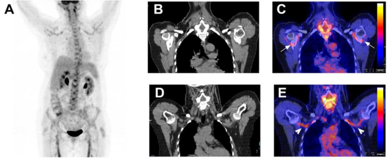
Figure 5.
A 64-year-old female patient with giant cell arteritis associated with polymyalgia rheumatica. A 64-year-old female patient was admitted with asthenia, headaches, shoulder, knee and elbow pain for 4 months. She had no fever. On physical examination, there was no sign of jaw claudication nor scalp tenderness. The temporal arteries were normal. White blood cells were normal. CRP was 35 mg/L. Temporal artery biopsy was negative. Coronal maximum intensity projection FDG-PET (A), coronal low-dose CT (B,D) and fused FDG-PET/CT (C,E) revealed FDG uptake in shoulders ((C) white arrowheads) and subclavian and axillary arteries ((E) white arrowheads). The diagnosis of giant cell arteritis associated with polymyalgia rheumatica was established. Her symptoms resolved with corticosteroids.

