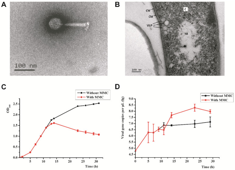
Figure 1.
Morphology and induction of PVJ1: (A) transmission electron micrographs of PVJ1; (B) thin section of a Psychrobacillus sp. GC2J1 cell following treatment with mitomycin C. CM, cytoplasmic membrane; NB, nuclear body; C, cytoplasm; CW, cell wall; VLP, virus-like particle; (C) effect of mitomycin C treatment on the growth of Psychrobacillus sp. GC2J1; (D) titer of PVJ1 during mitomycin C induction experiment.

