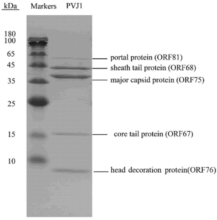
Figure 4.
Structural proteins of PVJ1 as revealed by SDS–PAGE. A sample of purified PVJ1 virions was subjected to 12% SDS–PAGE. The gel was stained with Coomassie brilliant blue G250. Gel slices containing protein bands were excised. Proteins in the gel slices were digested with trypsin, and the resulting peptides were analyzed by MALDI–TOF mass spectrometry. The identity of the proteins is shown.

