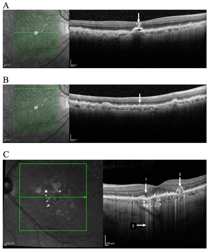
Figure 1.
Identification of OCT progression biomarkers of AMD in cross sectional SD-OCT scan. Example of (A) Hyperreflective foci (arrow); (B) Subretinal drusenoid deposits (arrow); (C) iRORA (1); Hyper-transmission defects (2); OCT-reflective drusen substructures (ODS) (3). Representative imagens acquired using OCT SPECTRALIS (Heidelberg Engineering, Heidelberg, Germany).

