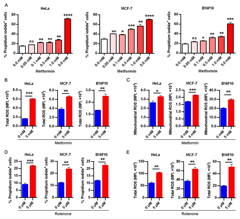
Figure 2.
Targeting oxidative phosphorylation by metformin promotes anoikis in cancer cells. (A) Apoptotic HeLa, MCF-7, and B16F10 expressed as percentage propidium iodide + cells treated with indicated doses of metformin in ECM detached condition for 48 h; (B) flow cytometry analysis of 24 h metformin treatment on total cellular reactive oxygen species (ROS) measured as mean fluorescence intensity in HeLa, MCF-7, and B16F10 stained with CM-H2DCFDA (5 μM) for 30 min; (C) flow cytometry mean fluorescence intensity quantification of mitochondria superoxide anion in HeLa, MCF-7, and B16F10 cells following metformin treatment for 24 h; (D) flow cytometry assay of percentage propidium iodide + HeLa, MCF-7, and B16F10 treated with specific complex 1 inhibitor-rotenone (5 μM); (E) flow cytometry analysis of 5 μM CM-H2DCFDA stained HeLa, MCF-7 and B16F10 presented as mean fluorescence intensity in 24hours treatment with rotenone All result are mean SEM, (n = 3) * p < 0.05, ** p < 0.01, *** p < 0.001 **** p < 0.0001, ns, not significant; typical data from at least three independent experiments with similar results.

