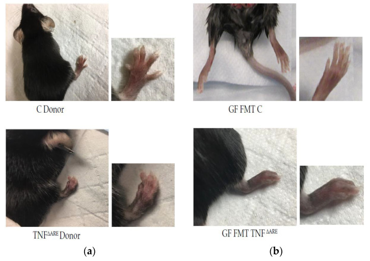
Figure 1.
Macroscopic in vivo phenotypes (a)—Representative pictures of donor mice. Top: male conventional donor control (C), n = 9. Bottom: male TNFΔARE+/− sick donor with enlargement of the right lower limb (n = 11). TNFΔARE+/− donor exhibits a degenerative arthritis with deformation and swelling phenotype compared to control. (b)—Picture of GF mice, that received FMT from C or TNFΔARE+/− donor. Top: control GF mice FMT with C donor which exhibit a phenotypically normal paw. Bottom: GF mice FMT with TNFΔARE+/− exhibiting a phenotype with deformation and swelling similar to the TNFΔARE+/− donor. Females show the same phenotypic pattern and pictures are representative of the entire colony. Histological examination of the gut and the joints of these GF male and female mice subjected to FMT with TNFΔARE+/− fecal matter, shows a degradation of the knee joints (clefts, debris, tears and loss of cartilage) and damages to the gut (thickening of the wall, disorganization of the villi, cell infiltration) compared to the group of male and female GF mice FMT with C mice, panels, see Figure 2a,c. Immunohistochemistry (IHC) was then conducted using anti-TNF antibody. In GF mice FMT with TNFΔARE+/−, as compared to GF mice FMT with healthy microbiome, IHC showed significant pro-inflammatory cytokine signature in tissues, particularly in the gut. We can localize the TNF staining in the gut epithelial layer cells and in the chondrocytes of the cartilage surface (Figure 2, panels (b,d) Results were similar between males and females).

