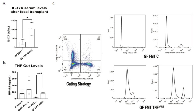
Figure 3.
Proinflammatory cytokines measurement and inflammation evaluation by flow cytometry. GraphPad Prism was used to generate the bar plots, that shows the mean of data and error bars show standard deviation. Stars represent significance (* for p < 0.05 and *** for p < 0.001). Multiplex ELISA Assay (Luminex). (a) Multiplex assay was conducted on the serum, with antibodies against TNF, IL-1β, IL-6 and IL-17. Results show a serological statistically significant (p = 0.0290) increase in IL-17A level in serum of GF mice FMT with TNFΔARE+/− donors (n = 6) compared to their controls, GF mice FMT with C donors (n = 3). No significant difference was detected in serum TNF, IL-1 β and IL-6 levels. Results were similar between males and females and were representative of the colony. (b) The multiplex assay was conducted at the tissue level after gut protein extraction. TNF levels were significatively elevated in TNFΔARE+/−donors (n = 4) compared to control donors (n = 4). TNF levels were below the limit f detection in healthy control mice donors. The levels of TNF were significatively elevated in GF mice FMT with TNFΔARE+/− (n = 4) compared to GF mice FMT with C (n = 3, p = 0.0004). (c)—Flow cytometry was conducted on spleen cells using antibodies directed against CD4 and CD8. After gating on lymphocytes, results show an activation and expansion of CD4+ and CD8+ T cells in GF mice FMT with TNFΔARE+/− when compared to their counterpart controls, suggesting an immune response initiation and inflammatory status initiation. Data shown from n = 4 in each group.

