FIG. 4.
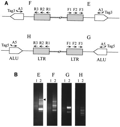
Detection of AMLV integrants by Alu PCR. (A) PCR amplification of virus-host junction fragments from AMLV-infected PBMCs was carried out using the primers described in Table 2 and indicated in the schematic diagram. (B) Panels E, F, G, and H show ethidium bromide-stained DNA fragments after electrophoresis on a 1.4% agarose gel of 10 μl of samples from the PCR reamplification of AMLV-infected PBMCs (lanes 2) or uninfected PBMCs (lanes 1). The primers used in panels E to H are indicated in the amplification strategies designated E to H, respectively, in the schematic diagram in panel A. Initially, 10 cycles of amplification were done using the Expand HF PCR system with 1.5 mM MgCl2 for AMLV F1-A3 (E) and AMLV F1-A5 (G) and 3.0 mM MgCl2 for AMLV R1-A3 (F) and AMLV R1-A5 (H). This step was followed by touchdown PCR for 40 cycles with AMLV F2-Tag3 (E), AMLV F2-Tag5 (G), AMLV R2-Tag3 (F), and AMLV R2-Tag5 (H). One microliter of the reaction was subjected to heminested PCR for 30 cycles using 2.5 U of Taq polymerase in PCR buffer containing 1.5 mM MgCl2 and AMLV F3-Tag3 (E), AMLV F3-Tag5 (G), AMLV R3-Tag3 (F) and AMLV R3-Tag5 (H). The arrowhead indicates the fragment that was isolated for nucleotide sequencing.

