Figure 5.
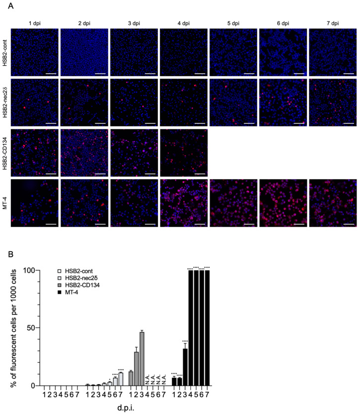
HHV-6B growth in HSB2-nec2δ cells. (A) Viral antigen detection in HSB2-cont, HSB2-nec2δ, HSB2-CD134, and MT-4 cells using an immunofluorescence assay. Cells were infected with the HHV-6B HST strain (multiplicity of infection = 0.1) and then treated with cold acetone for 1–7 days post-infection (d.p.i.). HHV-6 p41 protein (red) was detected in virus-infected HSB2-nec2δ and HSB2-CD134 cells. HSB2-cont cells were used as a negative control. HHV-6B-infected HSB2-CD134 cells died due to the lytic cycle at 4 d.p.i. Nuclei of cells were stained with DAPI (blue). Scale bars represent 50 μm. (B) Virus-infected cells (red) were counted 1–7 d.p.i. Error bars represent standard deviations (n = 3 per cell). Asterisks indicate statistical significances when compared with the HSB2-cont cells (* p < 0.05, **** p < 0.0001, Dunnett’s multiple comparison test except for the data of HSB2-CD134 because of missing values). N.A. indicates not applicable (uncountable due to lytic infection progression).

