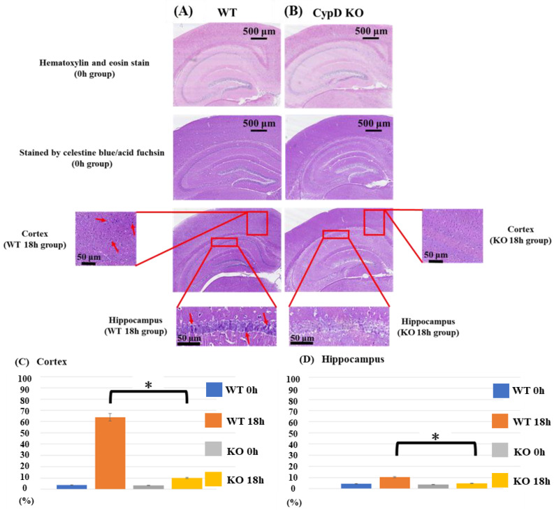
Figure 3.
Histological analysis. Images from acid-fuchsin and celestine blue-stained sections represent neuronal necrosis of cortex and hippocampal areas from WT (sham-operated control (A) and from CypD KO mice (B)). Neuronal damage in the cerebral cortex and the hippocampus was counted in a blinded manner and presented as the percentage number of damaged neurons (of the total number of cells). Tissues were harvested 18 h after surgery. Arrows indicate densely stained cells with high nuclear/cytoplasmic ratio, cellular swelling, and neuronal vacuolization. At 0 h after CLP treatment, there was no significant difference between the two groups regarding neither cerebral cortex nor hippocampus. At 18 h, neuronal cell death was significantly more severe in both the cerebral cortex and hippocampus in the WT group as compared to the KO group (C,D). Data are expressed as means ± SD, * p < 0.05, n = 5.

