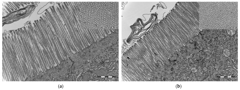
Figure 7.
Transmission electron microscopy micrographs from the midgut of cod larvae at 16 dph reared under (a) germ-free and (b) conventional conditions. Microvilli in conventional cod larvae were shorter and thinner compared to the microvilli in germ-free cod larvae. Insets in (a,b): Transverse section of brush border—microvilli were closer to each other in the conventional than in the germ-free cod larvae. Scale bars are 1 µm and of insets 500 nm.

