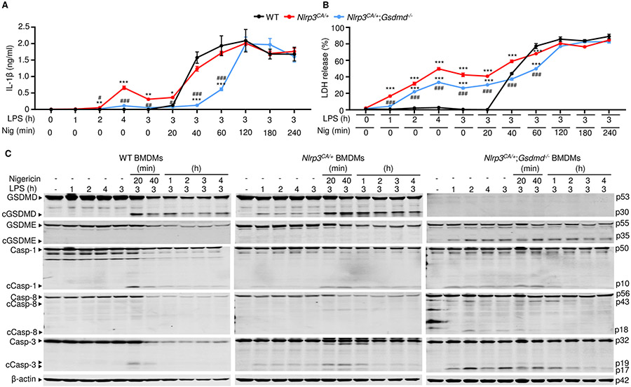
Figure 2. LPS stimulated IL-1β release and GSDME cleavage by Nlrp3CA/+;Gsdmd−/− BMDMs.
BMDMs were expanded in vitro in M-CSF-containing media from bone marrow cells isolated from WT, Nlrp3CA/+, or Nlrp3CA/+;Gsdmd−/− mice. BMDMs were primed with 100 ng/ml LPS for 1, 2, 3, or 4 hours and treated with 15 μM nigericin for 20 minutes, 40 minutes, 1, 2, 3, 4 hours. IL-1β (A) and LDH (B) in the conditioned media were measured by ELISA and by the cytotoxicity detection Kit, respectively. (C) The indicated proteins in the whole cell lysates were analyzed by immunoblotting. Data are mean ± SEM from experimental triplicates and are representative of at least three independent experiments. **P < 0.01; ***P < 0.001; ##P < 0.01; ###P < 0.001. **,***Nlrp3CA/+ or Nlrp3CA/+;Gsdmd−/− compared to WT; ##, ###Nlrp3CA/+;Gsdmd−/− compared to Nlrp3CA/+. One-Way ANOVA. BMDMs, bone marrow-derived macrophages; cCasp, cleaved caspase; cGSDM, cleaved gasdermin; h, hour; IL-1β, interleukin-1β; LDH, lactate dehydrogenase; LPS, lipopolysaccharide; min, minute; M-CSF, macrophage colony-stimulating factor; WT, wild type; CA, constitutive activation.

