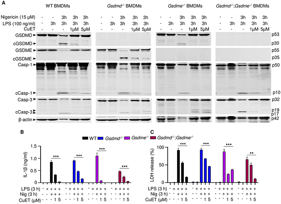
Figure 6. CuET inhibited GSDMD, GSDME, and IL-1β maturation and LDH release.
BMDMs were expanded in vitro in M-CSF-containing media from bone marrow cells isolated from WT, Gsdmd−/−, Gsdme−/−, or Gsdmd−/−;Gsdme−/− mice. BMDMs were primed with 100 ng/ml LPS for 3 hours and treated with vehicle or CuET for 1 hour before adding 15 μM nigericin for 3 hours. (A) The indicated proteins in the whole cell lysates were analyzed by immunoblotting. IL-1β (B) and LDH (C) in the conditioned media were measured by ELISA and by the cytotoxicity detection Kit, respectively. Data are mean ± SD from experimental triplicates and are representative of at least three independent experiments. **P < 0.01; ***P < 0.001. Two-Way ANOVA. BMDMs, bone marrow-derived macrophages; cCasp, cleaved caspase; cGSDM, cleaved gasdermin; h, hour; IL-1β, interleukin-1β; LDH, lactate dehydrogenase; LPS, lipopolysaccharide; min, minute; nig, nigericin; M-CSF, macrophage colony-stimulating factor; WT, wild type.

