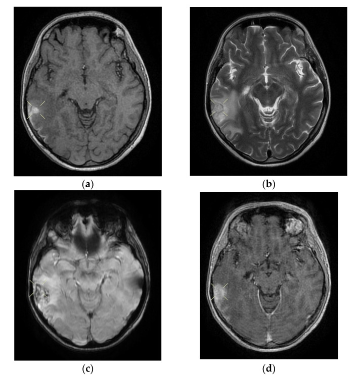
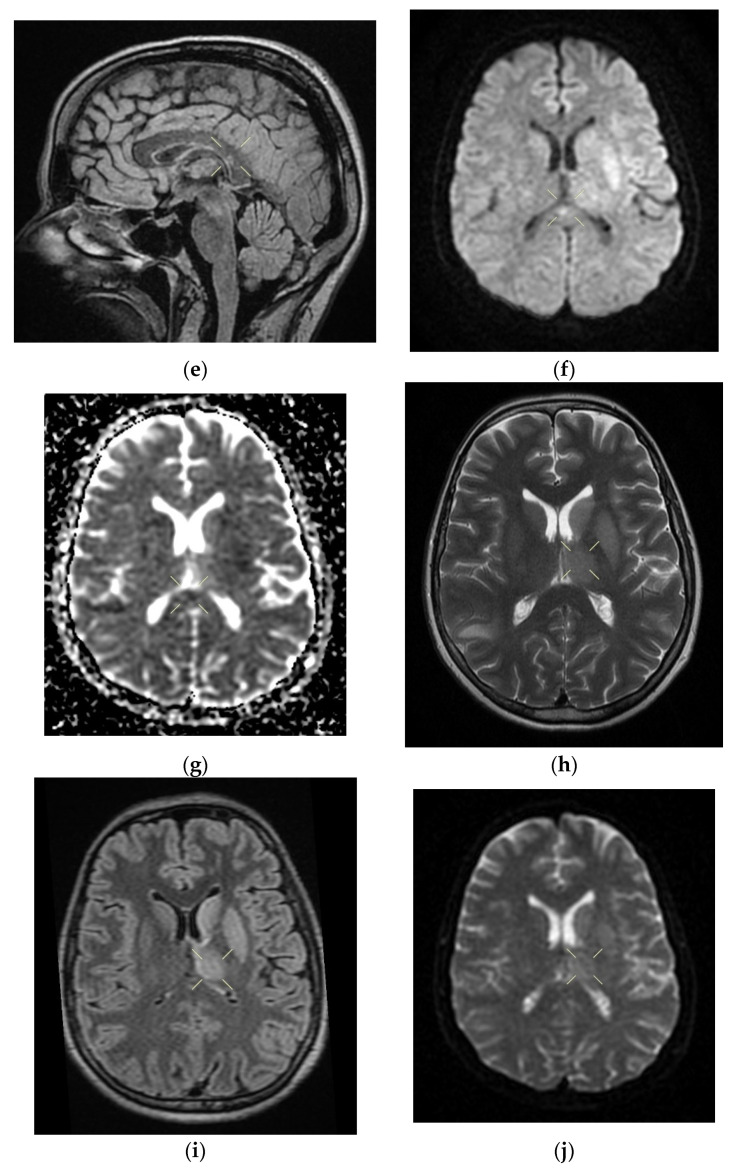
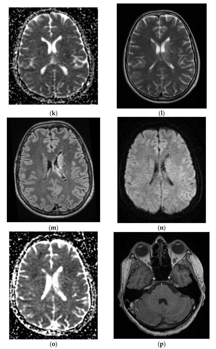
Figure 2.
Cerebral native and contrast-enhanced MRI and angiography, and CT cerebral venography highlighting the sigmoid sinus and right lateral sinus thrombosis and the inferior sagittal sinus and right sinus thrombosis, associated with right temporal cortical and subcortical subacute hemorrhage, supratentorial recent subacute synchronous lacunar infarct, (cytotoxic and vasogenic) thalamic–lenticular–caudal edema, and supratentorial non-specific demyelinating lesions. Magnetic resonance imaging shows cortico-subcortical subacute hemorrhage in the right temporal lobe (a,b) T1 and T2 hyperintensities. (c) methemoglobin signal. (d) heterogeneous contrast enhancement. (e) supratentorial recent subacute lacunar infarction in a millimeter lesion in hypersignal FLAIR, restrictive in diffusion coefficient. (f,g) supratentorial recent subacute lacunar infarction located in the corpus callosum. (h,i,j,k) cytotoxic and vasogenic edema in diffuse T2 and FLAIR high signal and moderate restriction in diffusion coefficient in the left thalamus. (l,m,n,o) cytotoxic and vasogenic edema in left lenticular-caudate nucleus. (p) right sigmoid and lateral sinuses thrombosis—T1 and T2 hyperintense material, without contrast enhancement. The intravenous post-contrast and native cranio-cerebral MRI examination highlights are as follows: oval globular formations with a non-homogeneous central portion and a periphery with a methemoglobin signal, hyper-intense T1–T2, axial dimensions of 11/10 mm maximally and heterogeneous contrast outlet, along with right temporal cortical and subcortical conglomerates, with extended moderate perilesional oedema; FLAIR hyper-intense millimeter lesions, intense and homogeneous restriction in diffusion and no-contrast outlets in the semioval centers, in the corpus callosum and in the middle temporal gyrus; diffuse signal T2–FLAIR increased in the left and left lenticular–caudal thalamus, with minimum diffusion restriction and no detectable contrast outlets; a few T2–FLAIR hyper-signal millimeter outbreaks, with no diffusion restriction and no corresponding T1, located in the white matter in the periventricular hemisphere and bilateral frontal–parietal subcortical area; normal supra- and infratentorial pericerebral liquid spaces; a symmetric ventricular system, with normal dimensions; structures of the median line in normal position; orbits and orbital content without anomalies; and paranasal sinuses with normal development and pneumatization. Magnetic resonance (MR) cerebral arteriography and venography indicated the following: internal carotid arteries symmetrically disposed, with a normal trajectory and caliber; anterior cerebral arteries and normal average bilaterally detached from the internal carotid, with no areas of stenosis or circumscribed dilation, with a homogeneous intralumenal signal; vertebral arteries, basilar artery, upper cerebral arteries and communicating arteries with a normal trajectory and caliber; hyper-intense T1–T2 material, with a no-contrast outlet, which transversely occupied the sinuses and sigmoid on the right side; and a lesion with the same signal characteristics situated along the right sinus and extended towards the inferior sagittal sinus; the rest of the dural sinuses had no detectable lesions in the sequences observed.


