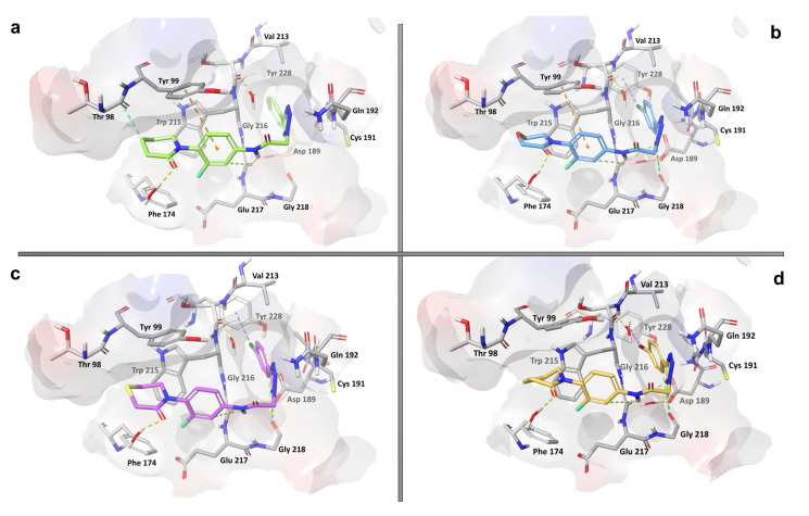
Figure 4.
Molecular docking poses of active peptide-1,2,3-triazole derivatives 8a, 8k, 8l and 8p (a–d) into the active site of the FXa enzyme (PDB: 2P16). The dotted lines indicate the most common ligand–protein interactions: H bonds in yellow, π–π stackings in orange, halogen–π interactions in magenta and aromatic H bonds in green.

