Figure 1.
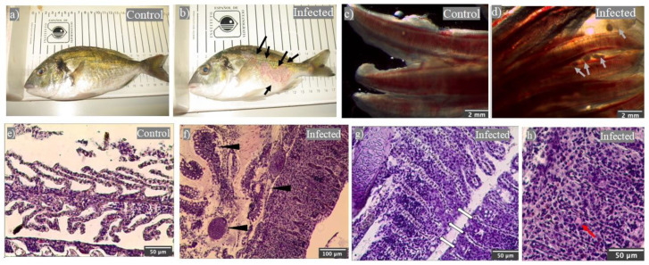
Cryptocaryon irritans trophonts are observed in the gills of gilthead seabream under a severe natural infection and produced histopathological alterations. Macroscopical representative images of healthy (Control: (a)) and C. irritans-infected (b) gilthead seabream. Binocular loupe representative images of gills from control (c) and C. irritans-infected (d) gilthead seabream. Histopathological study by light microscopy of gill sections stained with haematoxylin–eosin from control (e) and a C. irritans-infected (f–h) gilthead seabream. Note the parasite adhered to the gills (f). Scale bar: 2 mm (c,d) 50 μm (e,g,h), 100 μm (f). Black arrows point to skin injuries during C. irritans infection. Grey arrows and black arrow heads point to C. irritans trophonts. White arrows point to the undifferentiated tissue between the secondary lamellae. Red arrows point to eosinophilic cells.

