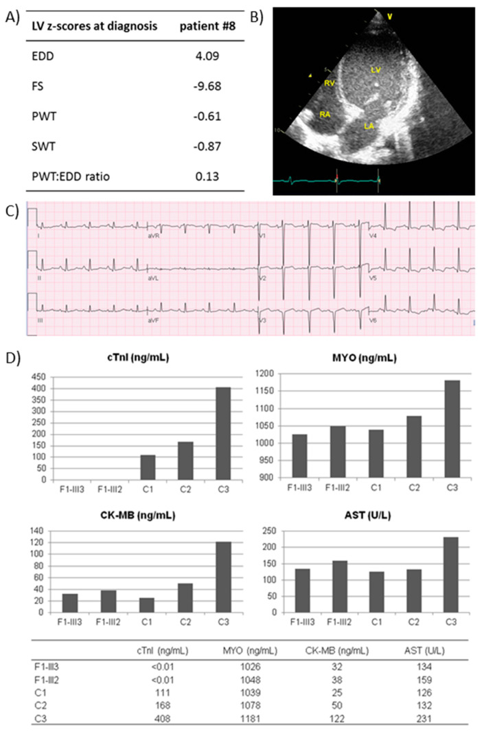
Figure 3.
Clinical presentation and cTnI tissue levels of patients with homozygous TNNI3 (OMIM *191044) variants. (A) Clinical data at admission of patient #8. (B) Two-dimensional echocardiogram of patient #8, showing a dilated left ventricle and left atrium, with rightward shift of the interatrial and interventricular septum and thin left free-walls. (C) Electrocardiogram of patient #8, showing low-voltage QRS complexes in inferior and lateral leads, no R wave progression in leads V1–V3 and T-wave inversion in leads V4–V6. (D) Histograms showing the tissue level of cTnI, myoglobin (MYO), muscular isoforms of creatine kinase (CK-MB) and aspartate aminotransferase (AST) in myocardial specimens from explanted frozen left ventricle of patient #41, her affected sister and three negative controls. F1-III3, patient #41; F1-III2, affected sister of patient #41; C1, age-matched hypertrophic cardiomyopathy patient; C2, age-matched idiopathic dilated cardiomyopathy patient; C3, adult ischemic cardiomyopathy patient. Abbreviations: LV, left ventricle; LA, left atrium; RV, right ventricle; RA, right atrium; EDD, end-diastolic dimension; FS, fractional shortening; PWT, posterior wall thickness; SWT, septal wall thickness.

