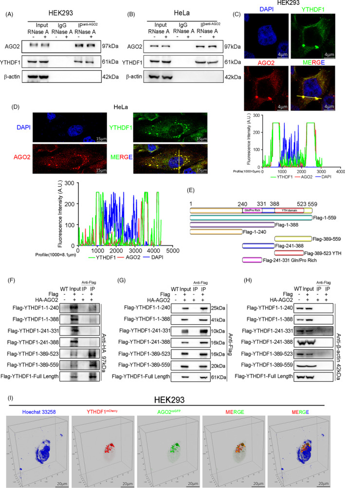
FIGURE 3.

YTHDF1 interacts with AGO2 through YTH domain. (A and B) Co‐immunoprecipitation (Co‐IP) and western blot had shown the binding of YTHDF1 and AGO2 in HEK293 (A) and HeLa (B) cell lines. (C and D) Immunofluorescence and laser confocal detection of co‐localization of YTHDF1 and AGO2 in HEK293 (C) and HeLa (D) cell lines. The graph below showed the fluorescence intensity peaks along the line. (E) Schematic diagram of human YTHDF1 and the fragments used in (F–H). (F–H) Co‐IP in lentivirus (YTHDF1 tagged with FLAG and AGO2 tagged with HA) infected HEK293 cells and western blot tested the HA (F), FLAG (G), and β‐actin (H). (I) Laser confocal images showed the co‐localization of YTHDF1mCherry and AGO2coGFP in vivo
