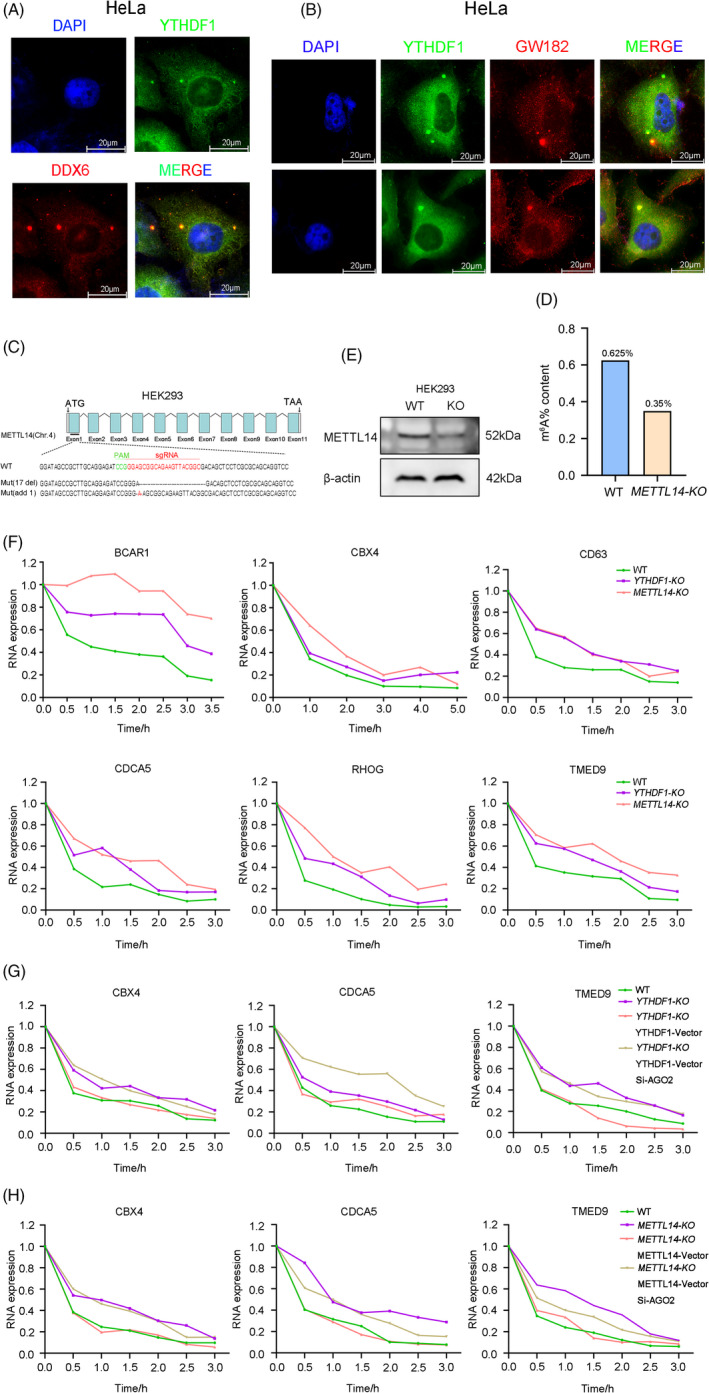
FIGURE 4.

YTHDF1 regulates mRNA degradation through AGO2. (A) Immunofluorescence imaging detected co‐localization of YTHDF1 and DDX6 in HeLa cells. (B) Immunofluorescence imaging detected co‐localization of YTHDF1 and GW182 in HeLa cells. (C) METTL14 was silenced by CRISPR‐Cas9 in HEK293 cells. Mutations in each allele were shown. (D) Content of m6A modification in HEK293 WT and METTL14‐KO cell lines. (E) Western blot analysis of METTL14 expresion in WT and METTL14‐KO HEK293 cell lines. (F) Half‐life change of the BCAR1, CBX4, CD63, CDCA5, RHOG, TMED9 by actinomycin D (10 μg/ml) inhibition in HEK293 cell lines (WT, YTHDF1‐KO, METTL14‐KO). (G) Half‐life change of the CBX4 CDCA5 TMED9 by actinomycin D (10 μg/ml) inhibition in HEK293 cell lines (WT, YTHDF1‐KO). YTHDF1‐Vector is represented overexpression of YTHDF1. Si‐AGO2 is represented knock down expression of AGO2 by siRNA. (H) Half‐life change of the CBX4, CDCA5, TMED9 by actinomycin D (10 μg/ml) inhibition in HEK293 cell lines (WT, METTL14‐KO). METTL14‐Vector is represented overexpression of METTL14
