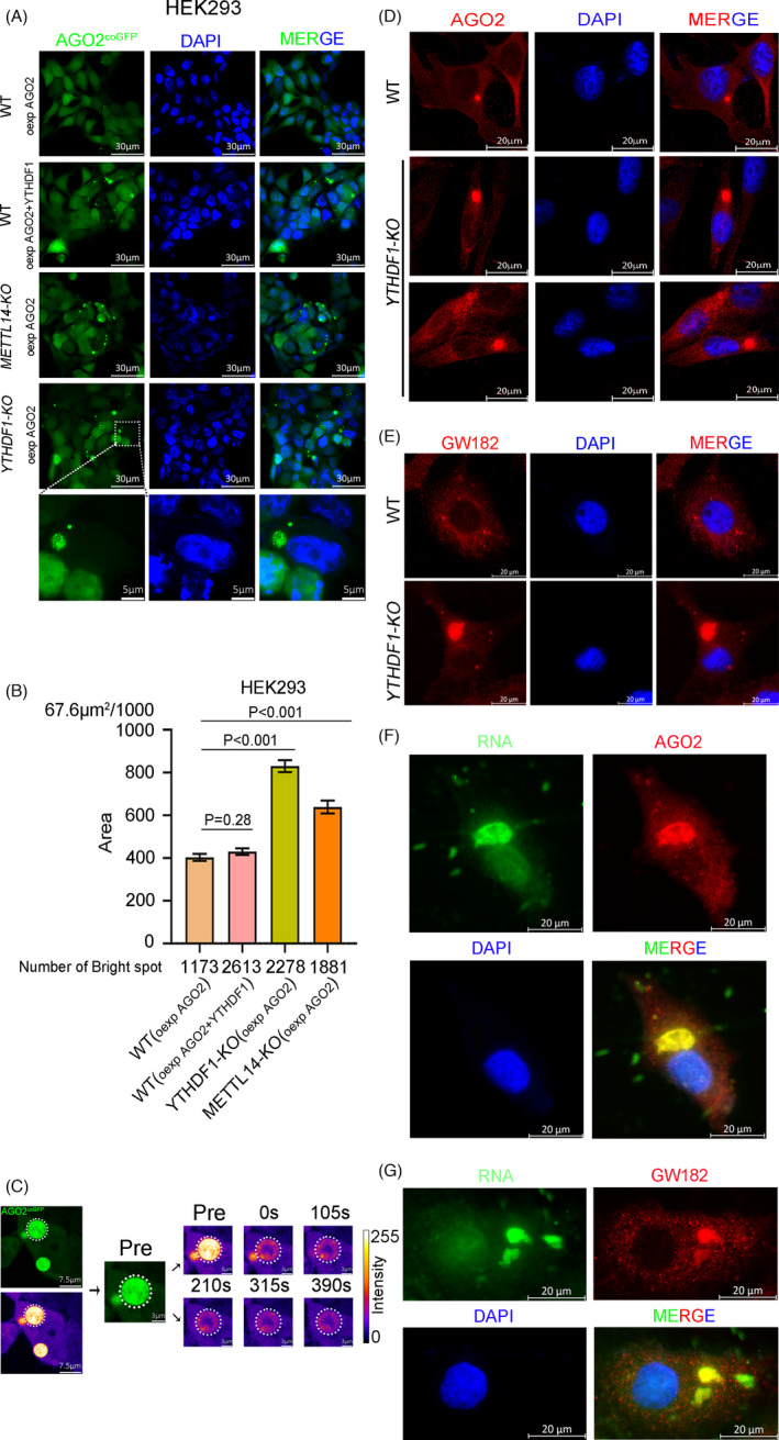
FIGURE 7.

Deletion of YTHDF1 impairs the P‐body formation. (A) Fluorescence microscope detection of AGO2coGFP foci in HEK293 cell lines (WT, YTHDF1‐KO, METTL14‐KO), “oexp” is represented overexpress. (B) Area of each AGO2coGFP foci in HEK293 cell lines (WT, YTHDF1‐KO, METTL14‐KO). (C) FRAP of partial photo bleached AGO2coGFP foci in YTHDF1‐KO HEK293 cells. (D and E) Immunofluorescence of AGO2 (D) and GW182 (E) in HeLa cell lines (WT and YTHDF1‐KO). (F and G) Fluorescence co‐staining of RNA dye and anti‐AGO2 (F)/GW182 (G) antibodies in YTHDF1‐KO HeLa cells
