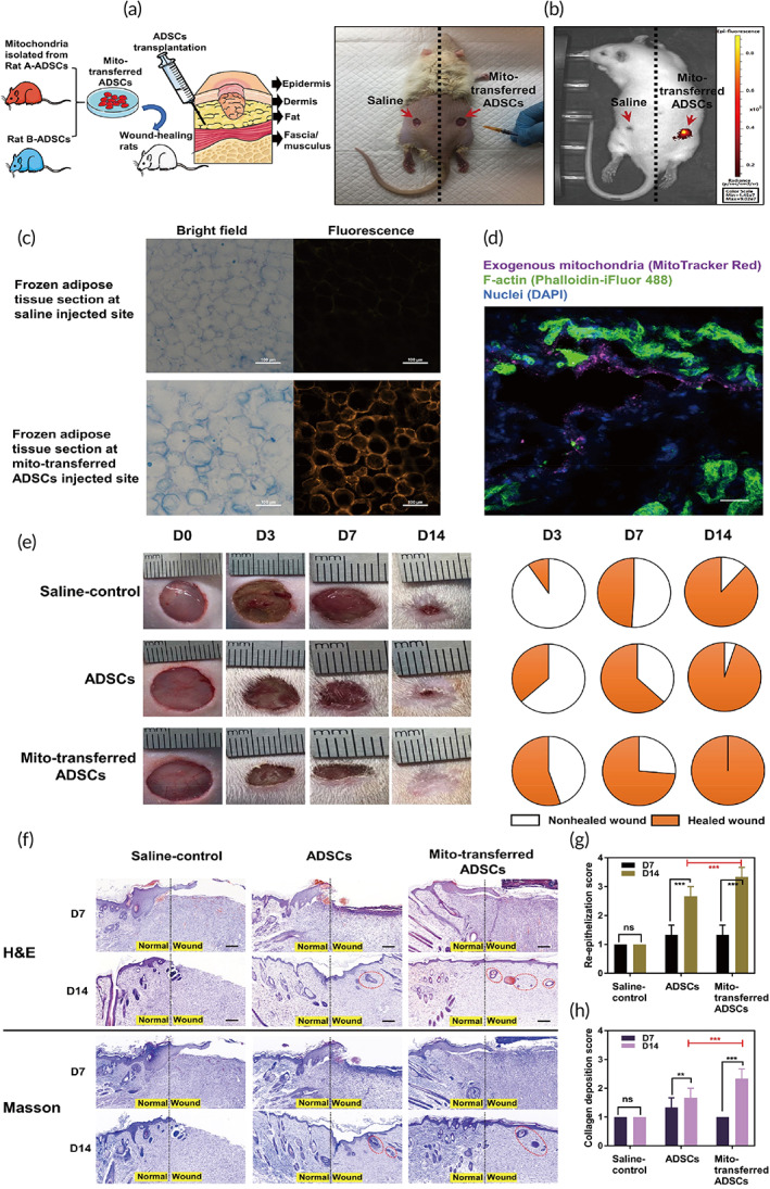
FIGURE 6.

In vivo assessment of mito‐transferred ADSCs in a rat full‐thickness skin wound model. (a) Schematic illustration of in vivo ADSCs injection into the fat layer of each wound. Each wound had about 10 mm in diameter and was 2 mm deep. The experimental groups are (i) sham control, (ii) rat B‐ADSCs, and (iii) mito‐transferred rat B‐ADSCs. (b) Tracking of infused mito‐transferred ADSCs by in vivo imaging. (c) The presence of fluorescence labeled exogenous mitochondria in adipose tissue. (d) Representative immunostaining images of adipocytes with exogenous mitochondria (MitoTracker Red), F‐actin (Phalloidin‐iFluor 488) and DAPI for nucleus. Scale bars, 20 μm. (e) Representative images of the wound area (left) and the corresponding fractions of wounds healed (right) by different treatments on Days 0, 3, 7, and 14 after operation (n = 8). (f) H&E and Masson staining of the wound area reflected the regenerated skin in different treatments at Days 7 and 12 (n = 3). Scale bars, 100 μm. New hair follicles were highlighted by red cycles. (g,h) The functional scores of re‐epithelization and collagen deposition was scored 0 to 4 (n = 8). Significantly different (one‐way ANOVA): ns, not significant, **p < 0.01, and ***p < 0.001
