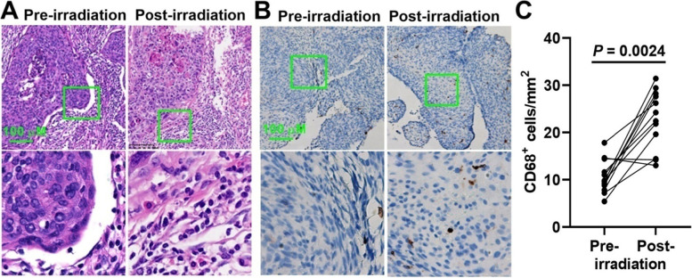
Fig. 1.
Immunohistochemical staining for tumor-associated macrophages (TAM) in cervical cancer tissue. The biopsies of cervical cancer patients were collected before and after radiotherapy. The cancer samples were fixed with formalin and embedded with paraffin. A, representative Hematoxylin-Eosin staining images. Images below were magnified 200×. B, representative images for CD68 staining. Images below were magnified 200×. C, the number of membranous CD68 positive cells was calculated in at least five randomly selected high power fields (400×). P value was calculated by Wilcoxon matched-pairs signed rank test

