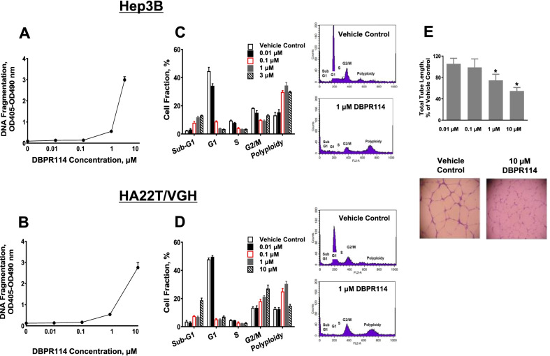
Fig. 1.
Effect of DBPR114 on apoptosis induction, cell cycle distribution, and HUVEC tube formation. A and B Apoptosis induction. Hep3B and HA22T/VGH cells were treated with DBPR114 at the indicated concentrations for 48 h in 10% FBS-containing cell culture medium. Apoptotic cell death was measured using DNA fragmentation ELISA. Mean ± SD, n = 3 replicates per concentration. C and D Cell cycle distribution. Cells were treated with DBPR114 in the presence of 10% FBS-containing cell culture medium for 48 h and then stained with propidium iodide. Cell cycle distribution was assessed using flow cytometry and quantified using FlowJo software. Mean ± SD, n = 3 replicates per concentration. Representative flow cytometry plots are presented for vehicle control and 1 μM DBPR114 from three replicates. E Tube formation of HUVEC. HUVEC were treated with DBPR114 at the indicated concentrations for 18 h. The tube formation was imaged, and the tube length was measured. Mean ± SD, n = 3 replicates per concentration. *p < 0.05 vs. vehicle control measured using unpaired t test

