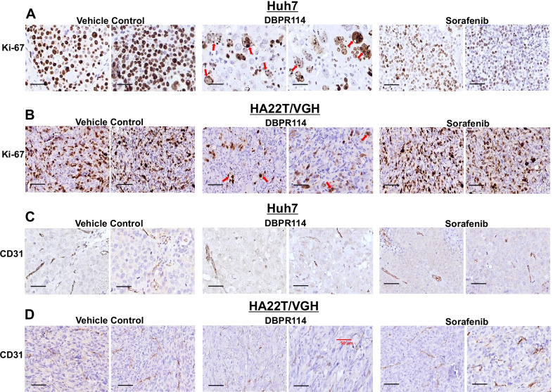
Fig. 5.
Histologic analysis of DBPR114 and sorafenib treatment in human HCC xenograft tumors. A and B Effect on cell proliferation (Ki-67 staining) and C and D effect on vascular endothelial cells (CD31 staining) in Huh7 and HA22T/VGH xenograft tumors. Tumor tissues were formalin-fixed and paraffin-embedded, with the paraffin sections used for immunohistochemical staining. Digital scans were performed with a 3DHITECH PANNORAMIC Midi slide scanner, and images were captured with PANNORAMIC Viewer software. Representative images were extracted from two separate animals in each group at × 40 magnification. Red arrows indicate apoptotic cell death and multinucleated cells. Bar: 50 µm

