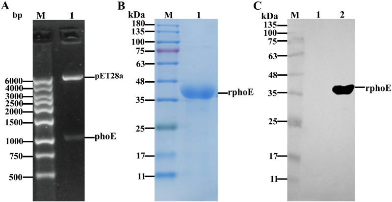
Figure 2.
Identification of pET28a-PhoE by restriction enzyme digestion and detection of purified PhoE by SDS-PAGE and Western blot analysis. A The recombinant plasmid was subjected to endonuclease digestion and identified by agarose gel electrophoresis; M: 1 kb DNA marker Ι, 1: the pET28a-PhoE plasmid was double digested with restriction endonucleases BamHI and XhoI. B SDS-PAGE analysis of purified PhoE protein (38.8 kDa). M: Pre-stained protein size maker; expected PhoE protein is indicated in lane 1. C. Western blot analysis of purified PhoE protein. The primary antibody was an anti-His tag monoclonal antibody, and the secondary antibody was an HRP-conjugated goat anti-mouse IgG. M: Pre-Stained Protein maker; the specific band of PhoE was present in lane 2, and lane 1 was the lysate of the strain carrying the empty pET28a vector.

