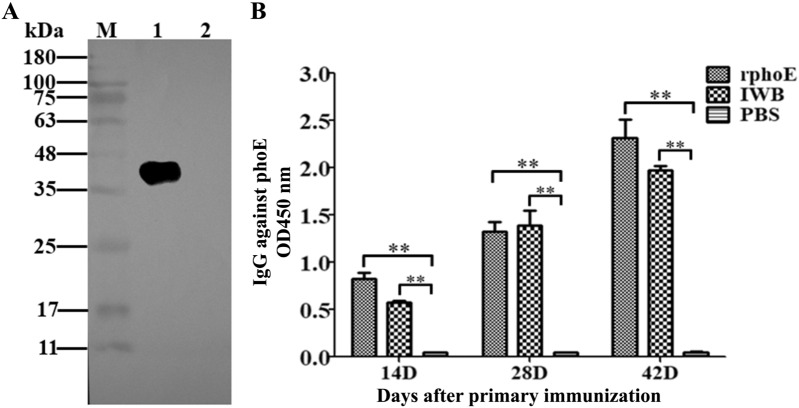
Figure 3.
PhoE-specific antibody responses in mice detected by Western blotting and iELISA. A Recognition of PhoE by mouse sera in a Western blot. Lane M: Pre-Stained Protein maker, lane 1 is the sample of purified PhoE protein; line 2 is the negative control of E. coli lysate containing the empty pET28a vector. For Western blot analysis, the primary antibody was the mouse serum isolated from the mice immunized with PhoE, the secondary antibody was an HRP-conjugated goat anti-mouse IgG. B Serum was tested for the presence of IgG antibodies by iELISA using PhoE as coating antigen. The absorbance of the developed HRP reaction was measured at 450 nm. Bars represent arithmetic means ± SD of antibody titers. **p < 0.01.

