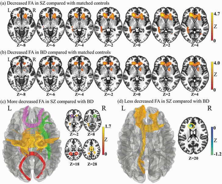
Fig. 1.
Whole brain meta-analysis of alterations in schizophrenia and bipolar disorder white matter fractional anisotropy. (a) Two-dimensional (2-D) representation of the significant clusters in the schizophrenia analysis. (b) Two-dimensional (2D) representation of the significant clusters in the bipolar disorder analysis. (c) Three-dimensional (3D) illustration of the more deceased FA in schizophrenia compared with bipolar disorder. The fibers of genu of corpus callosum are in purple, body in orange, and splenium in red. The green parts are the right Anterior limb of internal capsule. (d) Three-dimensional (3D) representation of the less deceased FA in schizophrenia compared with bipolar disorder. The yellow is the left anterior and superior corona radiata. The blue is the left external capsule extending to the posterior limb and Retrolenticular part of internal capsule and cerebral peduncle.

