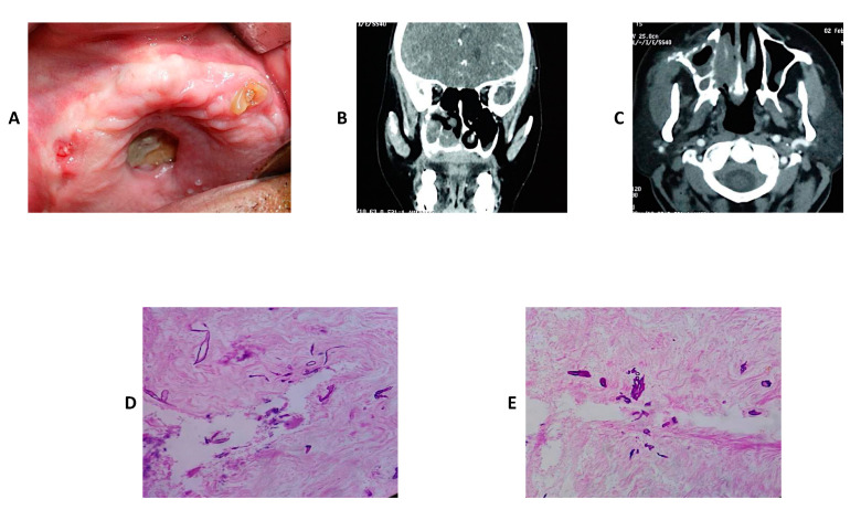
Figure 5.
An edentulous maxilla with an exposed necrotic bone in the palate and draining sinus in the right canine region (A). The axial (B) and coronal (C) slices of contrast enhanced CT scans of the maxilla respectively showing bone destruction, sequestrum formation, involvement of the right turbinate and sinus opacification. Histopathological slides (H&E staining) showing non-septate fungal hyphae consistent with diagnosis of mucormycosis (D,E).

