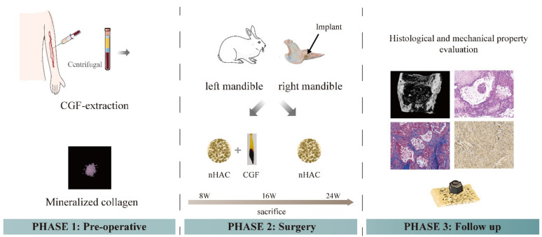
Figure 1.
Schematic diagram of the experimental process. The first step was the preparation process of CGF and its mixing with nHAC bone meal. The second step was the implantation process of animal experimental materials. Experimental group, nHAC/CGF; control group, nHAC. In the third step, the detection methods for the materials postoperatively included radiological examination, histological examination and biomechanical examination.

