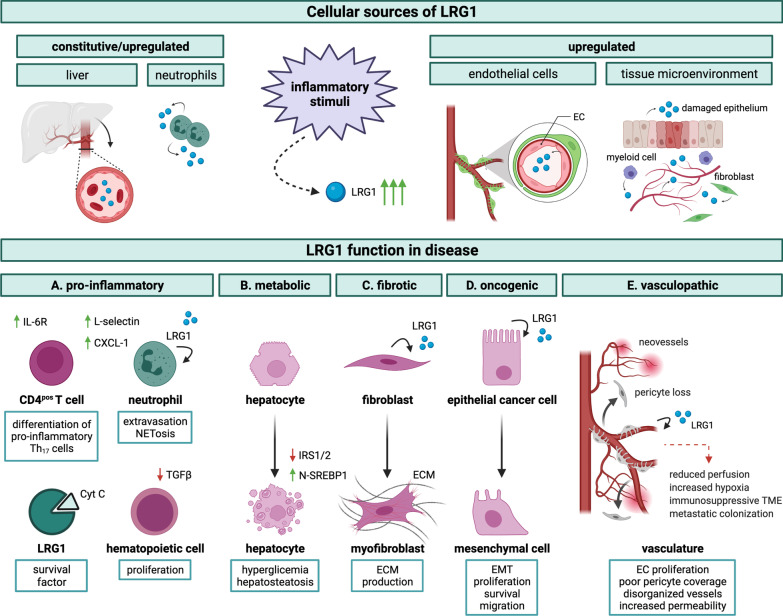
Fig. 5.
LRG1 functions in disease progression. Schematic representation of LRG1 cell sources and pathological functions. Following various inflammatory stimuli, including infection, injury, autoimmune disease, and tumour-associated inflammation, LRG1 may be produced systemically and/or at the local tissue level. Predominant cellular sources include hepatocytes, neutrophils, and endothelial cells but also other components of the tissue microenvironment, namely epithelial cells, fibroblasts, and other types of myeloid cells. LRG1 pathogenic functions may be initiated through autocrine and paracrine activity and can be broadly classified into A pro-inflammatory: LRG1 favours immune cell participation at the inflammatory site by (i) counteracting TGFβ-driven anti-proliferative function on hematopoietic progenitors; (ii) promoting the extravasation and activation of neutrophils; and (iii) enhancing the differentiation of naïve CD4pos T cells into pro-inflammatory Th17 lymphocytes. Additionally, LRG1 acts as a survival factor for circulating immune cells by neutralizing Cyt c cytotoxicity. B metabolic: LRG1 affects hepatocytes by suppressing fatty acid catabolism, promoting lipogenesis through activation of SREBP1, and inhibiting the expression of IRS1/2 thus contributing to hepatosteatosis and hyperglycemia. C fibrotic: LRG1 promotes the functional transition of fibroblasts (and epithelial cells, not shown) into ECM-producing cells in fibrosis. D oncogenic: LRG1 contributes to cancer cell malignancy by promoting EMT and exerting proliferative and anti-apoptotic functions. E vasculopathic: LRG1 affects vessel stability by promoting dysfunctional angiogenesis and interfering with EC-pericyte crosstalk. These effects contribute to the formation of disorganized and highly permeable capillaries. Notably, these outcomes indirectly sustain and amplify some of the direct effects, as dysfunctional and poorly perfused vessels are responsible for the establishment of a highly hypoxic microenvironment which, in turn, contributes to fibrosis, immunosuppression and cancer cell aggressiveness. Cyt c cytochrome c, IRS insulin receptor substrate, N-SREBP1 nuclear sterol regulatory element binding protein 1, ECM extracellular matrix, EMT epithelial-mesenchymal transition, EC endothelial cell

