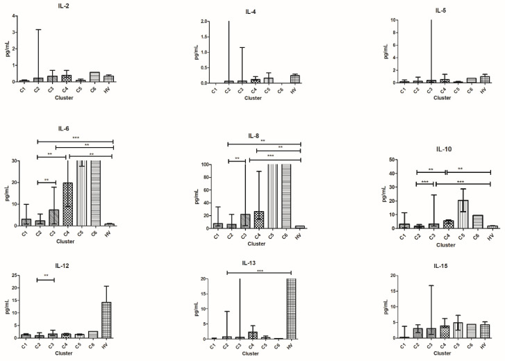
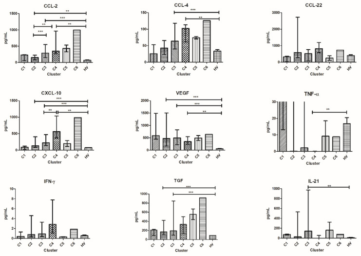
Figure 3.
Cytokine distribution at T0 in the six clusters and HV. Plasma concentration is expressed in pg/mL and plotted on the y-axis; clusters of patients are on the x-axis. For TNF-α, IL-4, IL-5, IL-6, IL-8, and IL-13 outliers are not shown for graphical choice. Data are expressed as median with range. *** p < 0.001, ** p < 0.01, * p < 0.05. CCL, C-C motif ligand; IL, interleukin.


