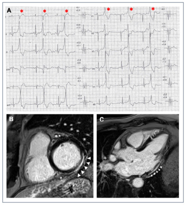
Figure 1.
A 40-year-old athlete presented with frequent PVBs on ECG ambulatory monitoring. Exercise test revealed frequent and repetitive monomorphic PVBs with RBBB/inferior axis morphology ((A), red asterisks). Post-contrast sequences on CMR showed an LGE subepicardial/midmyocardial stria in the basal inferolateral and inferior LV walls (white triangles; (B) short-axis view; (C) 3-chamber view).

