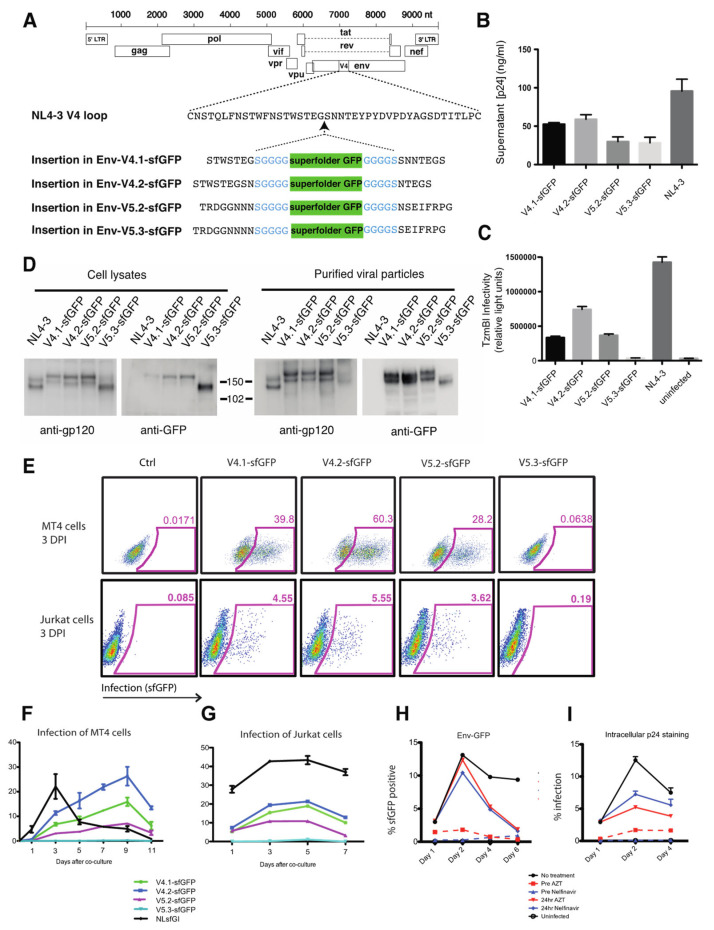
Figure 1.
Construction of infectious HIV clones with fluorescent Env carrying sfGFP inserted into V4 or V5 domains. (A) sfGFP is inserted into HIV-1(NL4-3) in V4 or V5. (B) Virus production by fluorescent Env HIV constructs following transfection of 293 T cells measured by p24 ELISA. (C) Cell-free virus infectivity was tested by infection of indicator cell line, Tzm-bl. Tzm-bl cells were infected with viral supernatants containing equivalent p24 antigen. (D) Western blot analysis of lysates of transfected 293 T cells or of virus particles harvested from transfected cell supernatants and purified through a 20% sucrose cushion. Blots were probed with anti-gp120 or anti-GFP antibody. Viral supernatants and cell lysates were collected at 48 h post transfection. (E) Infection of Jurkat cells or MT4 cells with virus was assessed on day 3 post infection. (F) Infection of MT4 cells initiated by co-culture with HIV-nucleofected Jurkat T cells. Flow cytometry was used to monitor the fraction of MT4 cells infected over time. (G) Infection of Jurkat cells initiated by spinnoculation of Jurkat T cells with cell-free virus. Flow cytometry was used to monitor the fraction of Jurkat cells infected over time. (H) Growth curve of HIV Env V4.2-sfGFP in Jurkat cells with and without treatment of AZT and Nelfinavir prior to or 24 h after infection measured by Env-GFP flow cytometry and (I) intracellular p24 staining.

