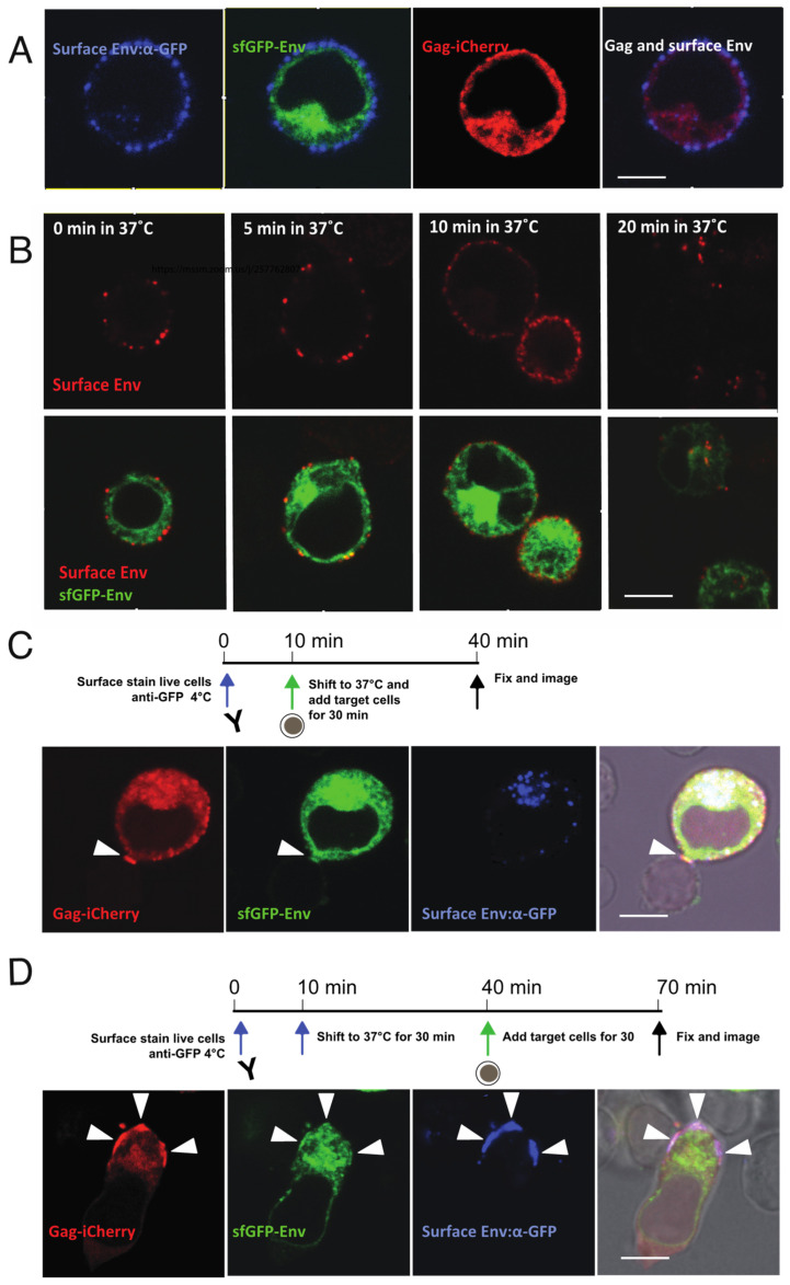
Figure 4.
Pulse-chase labeling of cell-surface Env illustrates internalization of Env and relocalization to VS. (A) Live cell surface staining of V4.2-Gag-iCherry: nucleofected Jurkat cells stained with anti-GFP antibody at 4 °C. (B) Pulse-chase of surface Env to determine time required for endocytosis: cells with surface-stained Env were shifted from 4 °C to 37 °C and kept for indicated time prior to fixation and imaging. (C) Stained cell according to the timeline was immediately co-cultured with primary CD4 target cells for 30 min at 37 °C and fixed for imaging. (D) Stained cell according to the timeline was first incubated at 37 °C for 30 min, then co-cultured with primary CD4 target cells for another 30 min at 37 °C and fixed for imaging. Arrowheads show VS. Bar: 6 µm.

