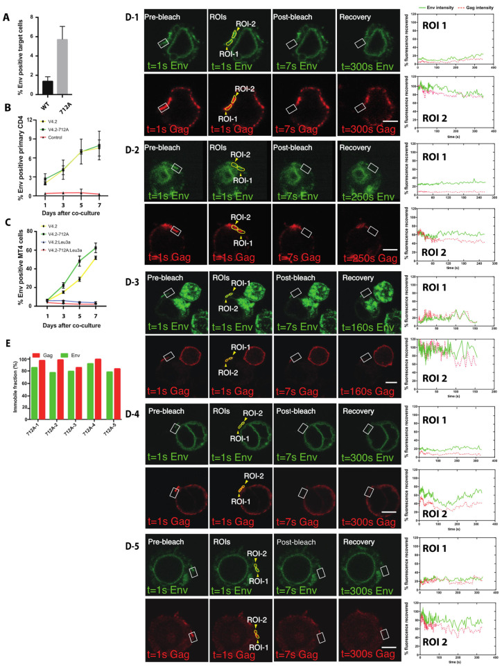
Figure 6.
Env fluorescence after photobleaching does not recover when examining Y712A mutants of Env V4.2-sfGFP in FRAP. (A) Jurkat cells nucleofected with wild type Env-V4.2-sfGFP or Env-V4.2-Y712A-sfGFP were co-cultured with primary CD4 cells for 3 h. Env transfer to primary CD4 cells were determined by Flow cytometry. (B) Jurkat cells nucleofected with wild type, Env-V4.2-sfGFP, or Env-V4.2-Y712A-sfGFP were co-cultured with activated primary CD4 cells to monitor productive infection in target cells. Samples were collected on day 1, 3, 5, 7 to determine the portion of primary CD4 cells with fluorescent Env. (C) Jurkat cells nucleofected with wild type, Env-V4.2-sfGFP, or Env-V4.2-Y712A-sfGFP were co-cultured with MT4 cells for days to monitor productive infection in target cells. Samples were collected on day 1, 3, 5, 7 to determine the portion of MT4 cells with fluorescent Env. (D) Fluorescence recovery after photobleaching (FRAP) of Env and Gag virological synapse with HIV V4.2-712A-Gag-iCherry. Before photobleaching a VS, both Gag and Env are concentrated at the junction between a donor cell and a target cell. A region of interest covering part of the synaptic button was bleached as shown in white square. ROIs were selected on bleached synapse (ROI-1) or an unbleached area (ROI-2) as shown in closed yellow region. Recovery curves of five individual experiments are displayed in (D-1–D-5). (E) shows the immobile fraction of each FRAP experiment. Bar: 5 µm.

