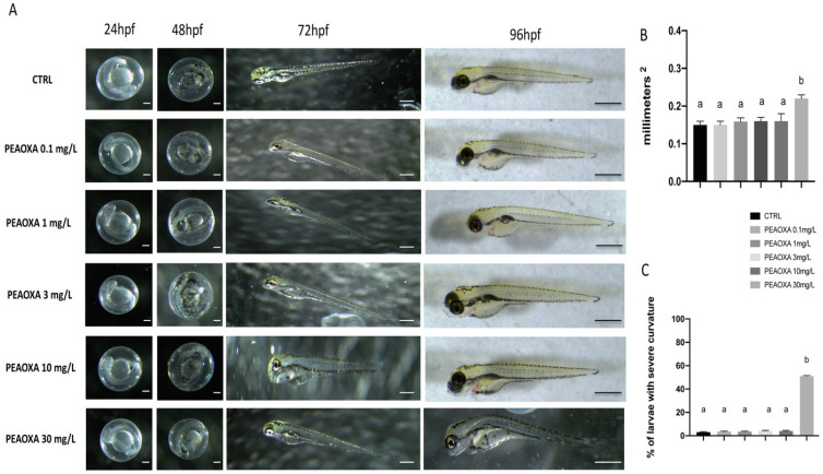
Figure 1.
The morphological abnormalities in zebrafish caused by PEAOXA different concentration exposure (A). Results for yolk sac area (B) and number of embryos with body axis curvature (C). Images were taken from the lateral view under a dissecting microscope (magnification 25). Scale bar, 500 μm. Values = means ± SD of three independent experiments Bars of group labelled with different letters (a, b) denote significant differences (p < 0.05).

