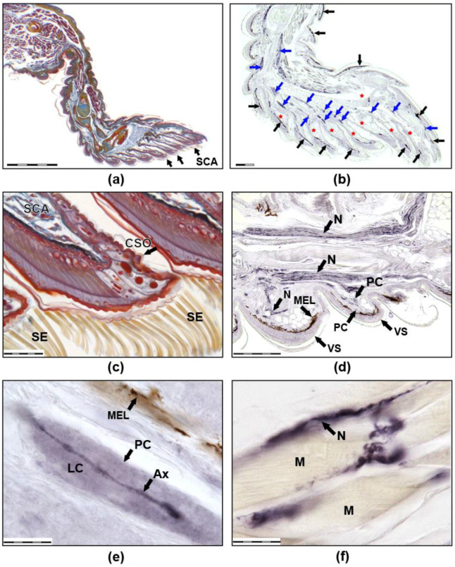
Figure 1.
Images (a,b) show sagittal sections of the third right anterior toe of a thick-toed gecko from SC group. (a) Mallory staining: the muscles are burgundy, the connective tissue is blue, the cartilage is yellow-blue, the bone is red, and the nerves are red-blue. (b) Immunohistochemical reaction with antibodies to NF 200. Positively coloured structures appear dark blue due to the use of NiCl2. Melanocytes look black-brown (not stained). Black arrows show melanocytes, blue arrows show nerves. Venous sinuses and their branches in the scansors are marked with red asterisks. (c) Distal parts of scansors, Mallory staining. Numerous setae on the ventral side of the scansor and CSO on the dorsal side of the scansor are visible. Images (d–f) show the immunohistochemical reaction with antibodies to NF 200 at higher magnification. (d) Large nerves are visible in the third phalanx of the toe and thinner nerves that innervate the ventral scales. (e) A single PC. (f) Muscle innervation. SCA: scansors or subdigital lamellae, SE: setae, CSO: cutaneous sense organ, VS: ventral scales, N: nerves, MEL: melanocytes, PC: Paciniform corpuscle, Ax: axone, LC: lamellar capsule, M: muscles). The size of the scale bars are as follows: (a) 500 mkm, (b,d) 200 mkm, (c) 50 mkm, and (e,f) 20 mkm.

