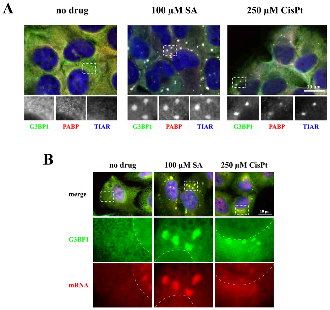
Fig. 2.

Detection of typical stress granules marker, poly(A)-binding protein (PABP), and mRNAs. (A) CisPt-induced foci contain PABP. One population of U2OS cells were used as unstressed control (no drug). Cells were stressed with sodium acetate (SA, 100 μM) or cisplatin (CisPt, 250 μM), for 1 h and 4 h, respectively. Then, cells were fixed and stained for G3BP1 (green), PABP (red) and TIAR (blue). All channels were demonstrated in grey in box region. The size bar represents 10 μm. (B) CisPt-induced foci do not contain mRNA. U2OS cells were treated with sodium arsenite (SA, 100 μM) for 1 h and cisplatin (CisPt, 250 μM) for 4 h (control cells, untreated, no drug). Cells were fixed and stained for G3BP1 and mRNA using FISH technique (G3BP1 – green – cyanine 2, mRNA – red – cyanine 3 fused with the anti-biotin secondary antibodies; in situ hybridization was done using oligo-dT40 probe against polyadenylated mRNA). Nuclei were visualized with Hoechst staining (blue). Boxed region is shown enlarged below each image, dotted line represents boundaries of nuclei.
