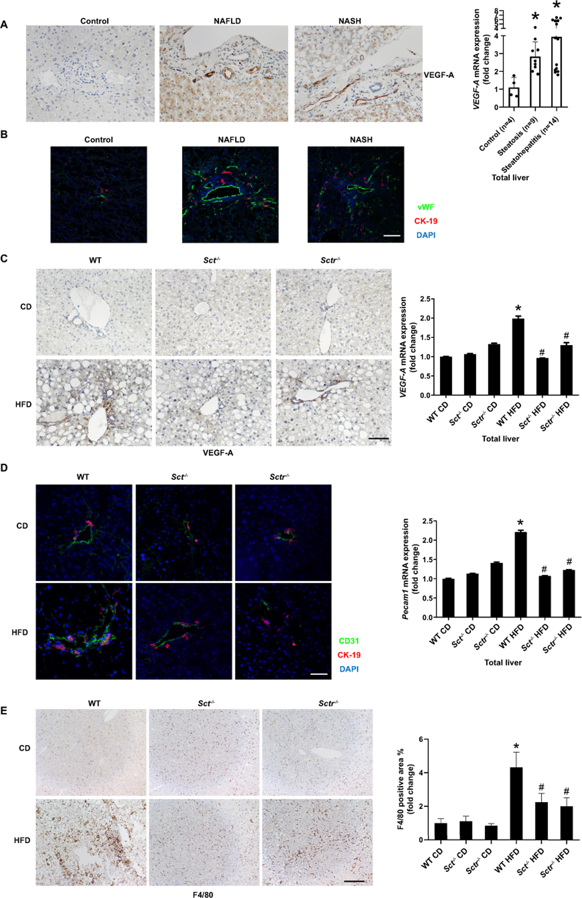
Figure 6. Knockout of the SCT/SCTR decreases HFD-induced liver angiogenesis and inflammation.

(A) VEGF-A expression in livers from NAFLD (n=9) and NASH (n=14) was measured by immunohistochemistry (20×; scale bar, 50 μm) and by qPCR. (B) Liver angiogenesis in liver samples from NAFLD (n=9) and NASH (n=14) was observed by immunofluorescence of vWF (20×; scale bar, 50 μm). (C) VEGF-A expression in Sct-/- and Sctr-/- HFD mouse liver was measured by immunohistochemistry (n=3; 20×; scale bar, 50 μm) and qPCR. (D) Angiogenesis in mouse liver samples was observed by immunofluorescence of CD31 (n=3; 40×; scale bar, 25 μm) and qPCR of Pecam1 in total liver (n=3). (E) Liver inflammation was detected by immunohistochemistry for F4/80 in Sct-/- and Sctr-/- HFD mice (n=24 from 8 different mice; 10×; scale bar, 100 μm). Data are mean ± SD. *P <0.05 versus WT CD or Control, #P <0.05 versus WT HFD.
