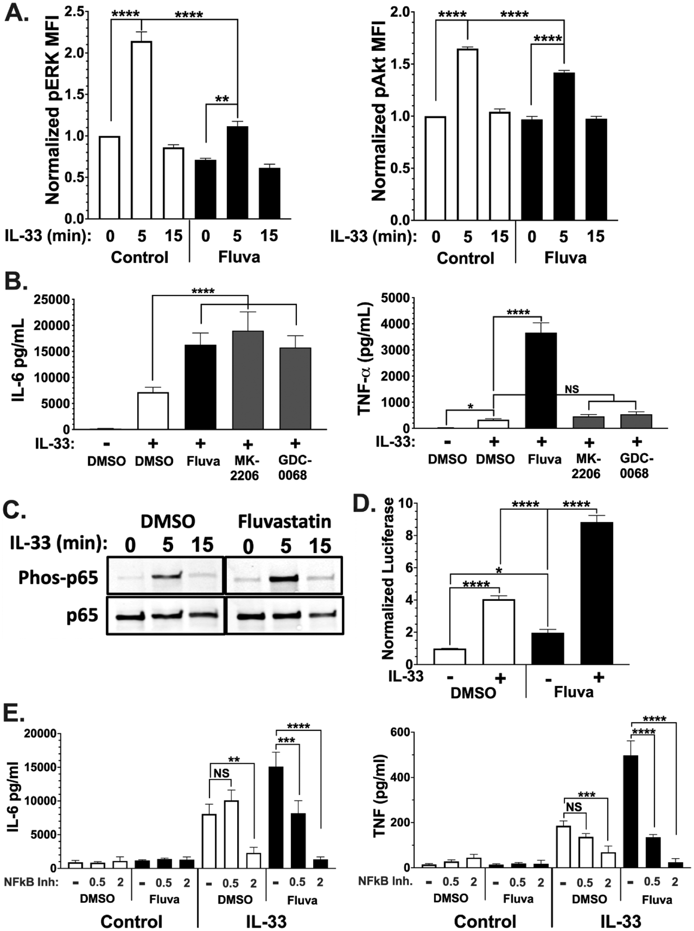
Figure 5. Fluvastatin alters IL-33 induced signaling.

(A) BMMC were cultured for 24 hrs in the presence of vehicle or fluvastatin (20 μM) followed by activation with IL-33 (100 ng/ml) for 0–15 minutes. Samples were fixed and stained with the indicated antibodies and analyzed via flow cytometry. MFI of phosphoprotein was normalized to vehicle 0' time point. Data shown are means ± SEM from 3 populations, representative of 2 experiments. (B) BMMC were cultured for 24 hours with the indicated chemicals prior to IL-33 stimulation for 16 hours. Culture supernatants were analyzed by ELISA. The Akt inhibitors MK-2206 and GDC-0068 were used at 3 μM and 5 μM, respectively. Data shown are means ± SEM from 3 populations. (C) Samples were lysed and analyzed by western blot. Data shown are from 1 of 3 experiments that yielded similar outcomes. (D) BMMC were transfected with vectors encoding luciferase genes from Renilla reniformis under HSV-TK promoter and Firefly under NF-κB response elements. Cultures were then treated vehicle or fluvastatin for 24 hours prior to activation IL-33 for 2 hours. Ratios of Firefly to Renilla luciferase were normalized to DMSO unstimulated. (E) BMMC were cultured in the presence of vehicle or fluvastatin ± bay11–7082. Cells were activated with IL-33 for 16 hours, and cytokines were measured by ELISA. Data are means ± SEM of 9 (D) or 8 (E) populations from 3 independent experiments.
