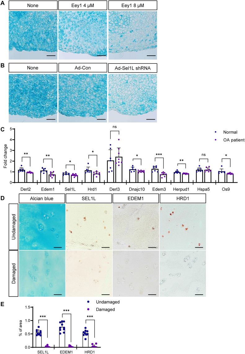
Fig. 5. Inhibition of ERAD function causes cartilage loss in mouse cartilage explants, and ERAD genes are down-regulated in OA chondrocytes.
(A) Mouse cartilage explants were treated with Eey1, and GAG deposition was visualized by Alcian blue staining. Eey1-treated explants displayed severe cartilage loss. Scale bars, 50 μm. (B) Sel1L expression in mouse cartilage explants was depleted by IA injection of adenoviral Sel1L-shRNA (Ad-sel1l shRNA), and GAG deposition was visualized by Alcian blue staining. Sel1L-depleted explants displayed severe cartilage loss. Scale bars, 50 μm. (C) Expression of ERAD genes in chondrocytes isolated from patients with OA was measured by qPCR. Most ERAD genes were down-regulated in OA chondrocytes. Normal, n = 7; OA patient, n = 8. (D) Immunohistochemistry (IHC) using several antibodies against ERAD genes showed decreased protein levels in OA cartilage tissue. Scale bars, 50 μm. (E) Measurement of SEL1L, EDEM, and HRD1 IHC density in OA cartilage tissues shown in (D). Undamaged, n = 10; damaged, n = 10. Statistical analysis was performed using the Student’s t test. ***(P < 0.001), **(0.001 < P < 0.01), *(0.01 < P < 0.05), ns (0.05 < P).

