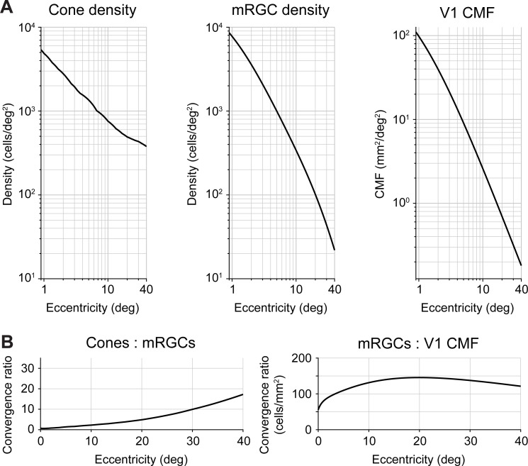
Fig 1. Foveal over-representation is amplified from cones to mRGCs to cortex.
(A) Cone density, mRGC receptive field density, and V1 cortical magnification factor as a function of eccentricity. Left panel: Cone data from Curcio et al. [9]. Middle panel: midget RGC RF density data from Watson [64]. Both cone and mRGC data are the average across cardinal retinal meridians of the left eye using the publicly available toolbox ISETBIO [65–67]. Right panel: V1 CMF is predicted by the areal equation published in Horton and Hoyt [68]. (B) Transformation ratios from cones to mRGCs and mRGCs to V1. The cone:mRGC ratio is unitless, as both cone density and mRGC density are quantified in cells/deg2. The increasing ratio indicates higher convergence of cone signals by the mRGCs. For mRGC:V1 CMF ratio units are defined in cells/mm2. The ratio increase in the first 20° indicates an amplification of the foveal over-representation in V1 compared to mRGCs.

