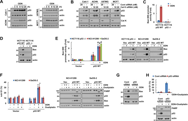
Fig. 3. Increased p53 expression contributes to ODN plus oxaliplatin-induced Bax upregulation and apoptosis.
A The cancer cell lines were treated with 2 μM ODN for the indicated times. B Cathepsin K-knockdown cell lines were used. C, D HCT116 p53 WT and p53-null cells were transiently transfected with a Bax/−600 promoter and incubated with 2 μM ODN for 12 h. The cells were lysed, and promoter activity and expression levels of Bax and p53 were measured. E The cancer cell lines were transiently cotransfected with vector or p53 WT and a Bax/−600 promoter and were treated with/without 2 μM ODN for 12 h, and promoter activity and expression levels of Bax and p53 were measured. F The cancer cell lines were transiently cotransfected with vector or p53 WT, and were treated with a combination of 25 μM oxaliplatin in the presence or absence of 2 μM ODN for 24 h. G, H Caki-1 cells were transfected with control siRNA or p53 siRNA, and treated with 2 μM ODN (G) or a combination of 2 μM ODN and 25 μM oxaliplatin (H) for 24 h. Protein expression and apoptosis were measured using western blotting (A, B, D, and E–H) and flow cytometry (F and H). The values in the graphs C, E, F, and H represent the mean ± SD of three independent experiments. *P < 0.01 compared to the ODN in HCT116 p53 WT cells. #P < 0.01 compared to the ODN in the vector-transfected cells. **P < 0.01 compared to the ODN plus oxaliplatin in the vector-transfected cells. †P < 0.01 compared to the control siRNA-transfected cells treated with ODN plus oxaliplatin.

