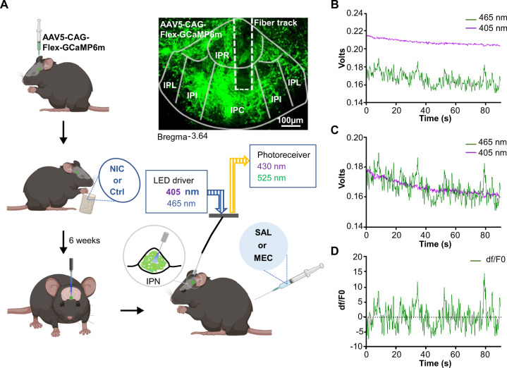
Fig. 1. Experimental design for combining GCaMP expression with fiber photometry to measure IPN GABAergic neuron activity during nicotine withdrawal.
A Experimental design for recording GCaMP activity from GAD2:Cre IPN neurons during nicotine withdrawal. AAV5 Cre-dependent GCaMP6m was injected into the IPN of GAD2:Cre mice. Mice then received nicotine or vehicle control solution in their drinking water for 6 weeks. Mice had optic fibers implanted into the IPN 4–6 weeks following viral injections. Calcium dependent (465 nm) and independent (405 nm) fluorescence was then recorded following IP administration of mecamylamine (1 mg/kg) or saline. For analysis of photometry data, demodulated fluorescence signals B of the 405 nm channel were scaled to the 465 nm channel (C) using least mean squares linear regression. The scaled signals were used to calculate the dF/F0 where dF/F0 = (465 nm signal – fitted 405 nm signal)/fitted 405 nm signal (D) that were used for subsequent analysis. NIC nicotine, Ctrl control, MEC mecamylamine, SAL saline, IPR interpeduncular rostral, IPL interpeduncular lateral, IPI interpeduncular intermediate, IPC interpeduncular caudal.

