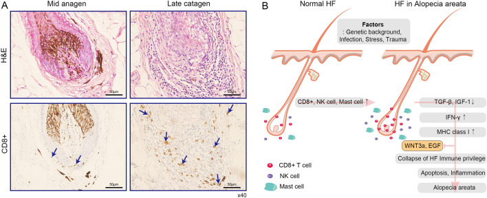
Fig. 6.
The interaction of perifollicular factors in AA. A H&E staining (top) and IHC (bottom) were performed with human anti-CD8 antibody in two different stages (left: mid anagen, right: late catagen) of biopsied HFs from AA patients. (Scale bar = 50 µm, ×40) A total nine individuals were tested (Fig. 6A and Fig. S1). Blue arrows indicate CD8+ T cells. B Schematic model for AA patient based on our results. Under normal conditions, immune cells such as CD8+ T cell, natural killer (NK) cells, and mast cells can be detected around HFs. Patients with a certain specific genetic background are predisposed to abnormalities in the micro-environment of the follicle. When various disruptions occur during anagen (e.g., infection, trauma, or stress), the clinical phenotype of alopecia areata results through above systemic mechanism (for more detailed explanation of this process, see the discussion section)

