Fig. 5.
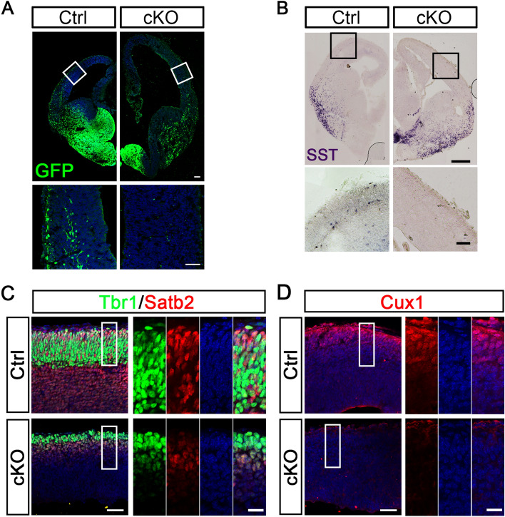
Congenital hydrocephalic mice exhibit abnormal neural development. A GFP staining of coronal sections from E14.5 control and cKO mice. GFP-labeled INs migrate tangentially into the cortex and form superficial and deep migratory streams in control embryos, whereas these streams are completely missing from cKO embryos. Boxes outline the regions shown below at higher magnification (scale bars, 100 μm for lower magnification; 50 μm for higher magnification). B Representative images showing in situ hybridization of SST mRNA expression in coronal sections from control and cKO at E14.5. Compared with controls, more SST-positive INs are generated in cKO embryos, but they are trapped at the ventral-dorsal boundary. Boxes indicate the regions shown below at higher magnification. SST-expressing cells normally migrate to the cortex in control embryos, but are almost absent from the cortex of cKO embryos (scale bars, 250 μm for lower magnification; 50 μm for higher magnification). C, D Tbr1, Satb2 (C), and Cux1 (D) immunostaining in the cortex of E14.5 embryos of control and cKO mice (scale bars, 50 μm for lower magnification; 20 μm for higher magnification).

