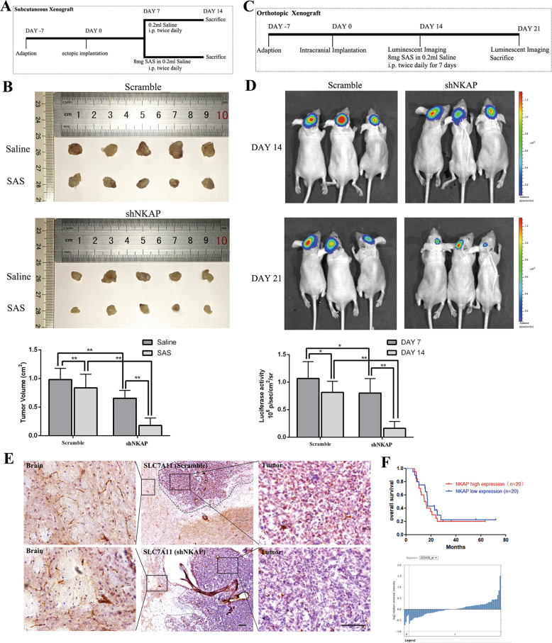
Fig. 4. NKAP suppressed glioblastoma growth and reduced sensitivity to ferroptosis inducer in vivo.
A Scheme showing the design of the subcutaneous xenograft model. B Representative subcutaneous xenograft tumor 14 days after inoculation showed reduced tumor volume and better efficacy of SAS in the shNKAP group. The bar graph indicates mean ± SD values of six independent animals. C Schematic showing the design of a subcutaneous xenograft model in an orthotopic intracranial mouse model. D Representative bioluminescence images of the intracranial tumor showed decreased luciferase activity and better efficacy of SAS in the shNKAP group. The bar graph indicates mean ± SD values of six independent animals. E Immunohistochemical labeling showed the downregulated expression of SLC7A11 in the shNKAP group. Scale bar = 100 µm. F Curves showed the better curative effect of standard therapy of glioblastoma patients in the NKAP high expression group. The graph below showed the relative NKAP expression level in 40 samples. *P < 0.05, **P < 0.01.

