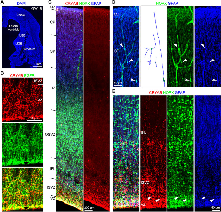
Fig. 3.
Identification of tRGs and oRGs in the developing human cortex. A A coronal section (60 μm thick) of the telencephalon including the cortex, LGE, MGE and striatum (caudate and putamen) of a 18 GW human fetal brain stained for DAPI. B CRYAB+EGFR+ tRGs in the cortical VZ. C HOPX, CRYAB and GFAP triple immunostained cortical section at GW18. Note HOPX+ cells in the cortical VZ, ISVZ, IFL, OSVZ, IZ, SP and CP, whereas CRYAB+ cell somas were mainly in the VZ. D HOPX+GFAP+ astrocyte lineage cells (arrowheads) in the cortical plate. E HOPX+CRYAB+GFAP+ tRGs (arrowheads) in the cortical VZ.

