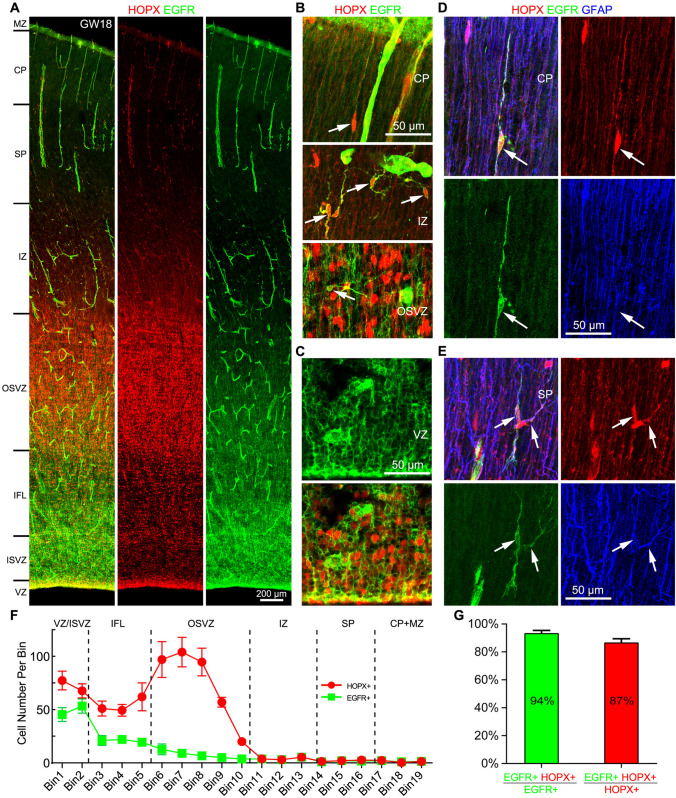
Fig. 5.
Human cortical tRGs and APCs express HOPX and EGFR. A HOPX and EGFR double immunostained GW18 human neocortical section. Note EGFR expression in blood vessels (pericytes and endothelial cells). B Higher magnification images showing HOPX+EGFR+ APCs (arrows) in the cortical CP, IZ and OSVZ. C HOPX+EGFR+ tRGs in the VZ. D, E HOPX+EGFR+GFAP+ APCs (arrows) in the cortical CP and SP. F Numbers of HOPX+ cells and EGFR+ cells in the cortex. Note that EGFR+ cells were mainly distributed in cortical VZ, ISVZ and IFL. G About 94% of EGFR+ cells expressed HOPX and 87% of HOPX+ cells expressed EGFR in the cortical IZ, SP and CP at GW18.

