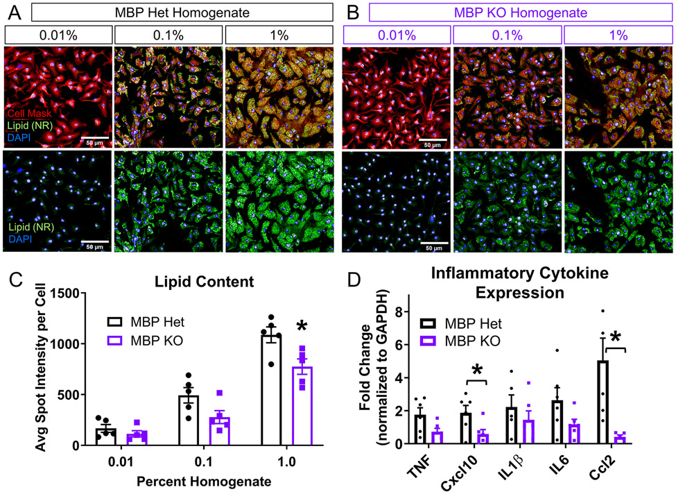
Figure 3. Foamy macrophages can form without myelin in vitro.
(A) Representative images from macrophages treated with 0.01, 0.1, and 1.0% (w/v) MBP Het homogenate, which contains normal amount of myelin. Neutral lipid droplets stained with Nile Red (green), cell membrane with Cell Mask (red), and nuclei with DAPI (blue), scale bar=50 μm. (B) Representative images from macrophages treated with 0.01, 0.1, and 1.0% (w/v) MBP KO homogenate, which is deficient in myelin. Neutral lipid droplets stained with Nile Red (green), cell membrane with Cell Mask (red), and nuclei with DAPI (blue), scale bar=50 μm. (C) Measurement of lipid droplet fluorescence demonstrates a concentration-dependent increase in intensity in macrophages after treatment with either MBP Het or KO spinal cord homogenate. Treatment with MBP KO homogenate showed decreased spot intensity compared to Het homogenate at 1% concentration (n=5 biological replicates, Two-way ANOVA with Bonferroni multiple comparisons, *P≤0.05 compared to MBP Het at corresponding concentration, error bars SEM.). (D) qPCR analysis of inflammatory cytokine expression (n=6 biological replicates per group, *p≤0.05 compared to MBP Het, Two-tailed Student’s t-test per cytokine, error bars=SEM)

